")
")
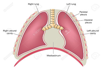
The lung is covered by a membrane called the pleura. The pleura is divided into the parietal and visceral pleura and between them there is always a small amount of fluid (1-2ml). In pathological situations the fluid increases.
The presence of this fluid could be found randomly during radiology examination or the patient may be experiencing some symptoms, for example shortness of breath, cough and chest pain.
Different diseases may cause an increase in the fluid for example heart failure, cancer, infections, Tuberculosis, autoimmune diseases and trauma.
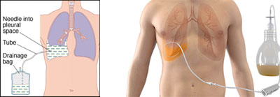
The initial approach of these patients is done via thorococentesis. In this procedure a fine needle is inserted into the chest wall to collect a small amount of fluid for testing.
In some cases the fluid is too large and must be drained or a chest drain may be required for a few days, until the fluid is removed.
When the fluid results are inconclusive, then we may sometimes proceed to thoracoscopy.
The treatment of the fluid is dependent on the cause.
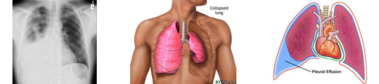
PNEUMOTHORAX
This is a condition defined by the presence of air between the visceral and parietal pleura, which leads to the collapse or partial collapse of the lung.

Pneumothorax depending on the cause could be spontaneous, traumatic or iatrogenic.
The symptoms of a patient with a pneumothorax irrespective of the cause are identified by sudden chest pain, shortness of breath and cough. It is also common for the patient to have tachycardia, tachypnoea, and oxygen desaturation. A life threatening complication of pneumothorax is tension pneumothorax. This affects the heart function and needs immediate intervention.
The treatment of pneumothorax aims at re-expanding the lung, which could be achieved either by oxygen administration or by placement of a chest drain.